A small pouch placed under the liver, which is essential for the digestive process, may be affected by the formation of stones. It is the gallbladder or cholecyst, where the bile produced by the liver is collected before being transported to the intestine where it will participate in fat digestion. The risks associated with the presence of gallstones and the role of endoscopy in diagnosis and treatment are described by Dr. Silvia Carrara, Head of the Humanitas Ecoendoscopy Programme.
Who is most at risk?
Cholecystic stones are formations of various sizes and color composed of different substances such as fats, cholesterol, bile salts or bilirubin. The latter is a substance that contributes to the formation of bile and it is the result of the degradation of hemoglobin. Its color is reddish-yellow and when the calcifications are composed of bilirubin they assume a darker pigmentation.


“The reason why gallstones develop is not yet clear. However – explains Dr. Carrara – many people have a high concentration of cholesterol and/or calcium inside the bile (above average) from which calculations can be developed”.
There are several factors that increase the risk of developing gallstones:
- Sex: they are more common in women;
- Age: the risk of gallstones increases with increasing age, being extremely rare in children and progressively more frequent over time, especially after the age of forty;
- Family history: gallstones are more common in some families, suggesting that genetics could play a role in the development of the disease.
Other conditions that may increase the risk of developing gallstones are: pregnancy, medications containing estrogen (such as contraceptive pills), obesity, frequent and prolonged fasting, rapid weight loss (including patients who have undergone surgical treatments for weight loss), lack of physical activity, diabetes mellitus, sickle cell anemia (and other conditions associated with rapid destruction of red blood cells, such as in patients with mechanical heart valves)”.
The pancreas is also involved
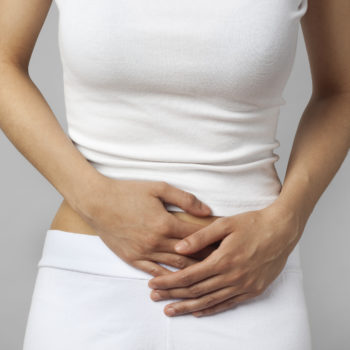
The presence of calcifications is not always symptomatic. Sometimes they may be found by chance when radiological examinations of the abdomen are carried out for other reasons. The patient has symptoms when the gallstones block bile ducts, the path of passage of the bile to the gallbladder. Abdominal pain, nausea and vomiting are the most frequent symptoms. Pain from the upper abdomen can also radiate to the back and it may be accompanied by fever.
In some cases, gallstones may also hinder the passage of digestive enzymes from the pancreas, leading to inflammation of the pancreas called pancreatitis. Another possible consequence of the presence of gallstones is cholangitis, i.e. inflammation of the bile tract for bile retention. The manifestation of jaundice is a suggestive symptom of cholangitis.
How to eliminate gallstones
The treatment of the calcifications is preferably surgical if these small pebbles are found in the cholecyst. A cholecistectomy is usually performed in laparoscopy (in some complex cases it is possible to resort to classical intervention in laparotomy, with open abdomen), with several small incisions of the abdominal wall, a minimally invasive practice. Drug therapy to dissolve cholesterol calcifications is a possible option but it takes a long time and once discontinued the risk of recurrence is high.
What is the role of endoscopy in the diagnosis and treatment of gallstones? “The diagnostic role of endoscopy, and more precisely the ecoendoscopy (EUS), is complementary to classical diagnostic imaging techniques (abdomen ecography, magnetic resonance imaging) for searching for gallbladder stones. Ecoendoscopy plays a major role, together with magnetic resonance, in the diagnosis of calcification of the main biliary pathway, which represents a clinical condition that is often symptomatic and causes biliary colic type pain, largely associated with the presence of gallstones”.
Endoscopic treatment of the main biliary route calcifications is performed through a further procedure, ERCP (endoscopic retrograde colangio-pancreatography). With this method, through the use of an endoscopic probe and some catheters dedicated to the procedure, the bile calcifications are removed after having made a small cut of the papilla (sfinterotomy), which represents the outlet of the bile route in the duodenum”.
Inflammation and cancer
Are gallstones episodes related to cancer risk? “The association between cholecystic tumors and calcifications, as well as the cause of calcifications, has not yet been demonstrated. However, the presence of calcifications was noticed in almost all cases of cholecystic cancer. Calcifications may cause chronic inflammation within the cholecyst, leading to the appearance of dysplasia (changes in cells) and subsequent cancer “.
The risk could also increase the more frequent episodes of cholecystitis, mostly related to the presence of calcifications. At the moment, there is no justification for the need to remove the gallbladder beforehand in patients who have calcifications that do not cause symptoms, as the risk of developing cancer in these patients is very low,” concludes Dr. Carrara.